Back to Blog

MRIs show: If you don’t move it, you lose it!
I just happened to check out Discovery News Wednesday to see that there was a new article published in this month’s Neurology Journal.
The study looked at 10 right hand dominant subjects that had sustained a right sided upper extremity injury that required immobilization. Each subject had an MRI within the first 48 hours of the injury, as well as one after 14 days of immobilization:
Exerpt: “The entire right arm was immobilized, preventing even small movements in the arm and hand.”
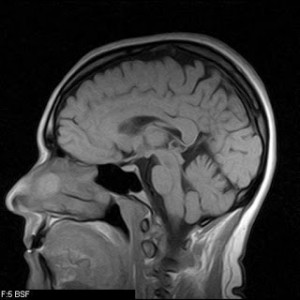
Comparing the initial MRI to the 2 week follow-up, there was a decrease in the cortical thickness in the primary motor areas of the brain designating to the right arm and hand suggesting complete disuse of the limb will create plastic changes in the brain.

Interestingly the area of the brain that controls motor function of the left upper extremity (the patient’s non-dominant side) had increased in size, since most of these patients were required to now use the left arm for tasks such as feeding and brushing their teeth. Over the two weeks these patients became more dexterous with the left hand (objectified with a series of fine motor tests), and these changes can be seen objectively on MRI.
Although this study shows that extreme immobilization can result in plastic changes of the brain, one can apply this to patients who have been cast at a single joint. This also sparks the question that if you are generally inactive will that result in general loss of brain volume in motor areas?
The study by N. Langer, MSc and L. Jancke will also be following these patients after removal of the immobilization device. How long will it take for the cortical area to expand to pre-injury size, or will it achieve this thickness again?
N. Langer, et. al. (2012). Effects of limb immobilization on brain plasticity. Neurology. 78, 182-188.

